We study Group B streptococcus (GBS)
We are interested in understanding fundamental mechanisms of bacterial pathogenesis and cellular innate immune/inflammatory defense responses during infection. GBS, also known as Streptococcus agalactiae, is a Gram-positive, β-hemolytic opportunistic pathogen that is the leading cause of bacterial meningitis in newborns as well as invasive infections in pregnant or immunocompromised individuals.
Currently there is no vaccine available to prevent GBS disease; guidelines focus on a single strategy of screening for GBS vaginal carriage during pregnancy with intrapartum antibiotic prophylaxis for all carriers. However, these strategies have not prevented GBS neonatal sepsis and meningitis and there is increased concern of emerging patterns of antibiotic resistance of other bacteria present during treatment as well as clindamycin resistance in GBS. Additionally, the incidence of GBS disease in non-pregnant adults is increasing worldwide with diabetes being a leading comorbidity.
Our research focuses on elucidating the host-pathogen interactions that promote GBS colonization and disease with the goal to inform new treatment or prevention strategies.
Research Areas
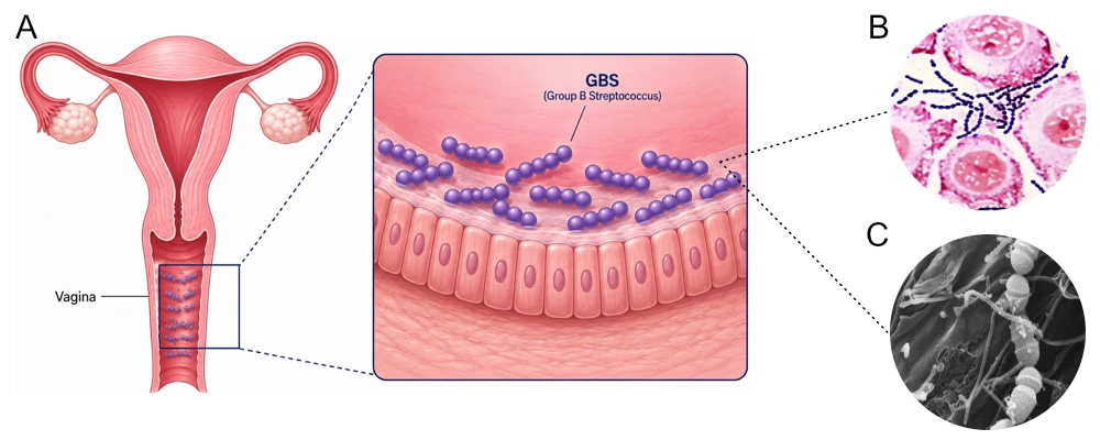
Figure (A) illustrates GBS vaginal colonization. Figure (B) is a Gram stain of GBS attaching to human vaginal cells. Figure (C) is an SEM of GBS attaching to human vaginal cells.
GBS colonizes the gastrointestinal (GI) tract and the vaginal tract of ~30% of individuals. During pregnancy, GBS can be transmitted vertically from a colonized mother to the newborn in utero or during childbirth, which frequently causes early-onset neonatal invasive disease (EOD) such as sepsis and pneumonia in the first week of life. Thus, maternal vaginal carriage is an important risk factor for newborn disease.
Our studies seek to characterize the bacterial and host factors as well as microbiome interactions that promote GBS vaginal persistence and accession to the uterus. We use a murine model of vaginal colonization, human cell lines as well as human isolates to study mechanistic interactions and immune responses.
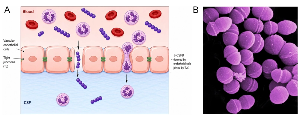
Figure (A) illustrates the pathogenesis of GBS Meningitis. Figure (B) is an SEM of GBS strain isolated from a newborn with meningitis.
GBS is the leading cause of neonatal meningitis, which can result in mortality rates of up to 9% and long-lasting neurological sequelae in survivors. GBS meningitis typically presents after 1 week of life and is classified as late-onset disease (LOD), which accounts for approximately 60% of GBS meningitis cases reported. GBS LOD is thought to occur following colonization of the neonatal GI tract, which can result from ingestion of infected amniotic fluid in utero, vaginal fluid during childbirth, or infected breast milk postnatally.
We use a neonatal murine model of GBS LOD to study GI colonization, dissemination to the brain and the inflammatory response that promotes meningitis. We seek to identify bacterial factors that promote penetration of gut and brain barriers as well as how host factors, such as mucins, and immune cells, such as neutrophils, contribute to pathogenesis
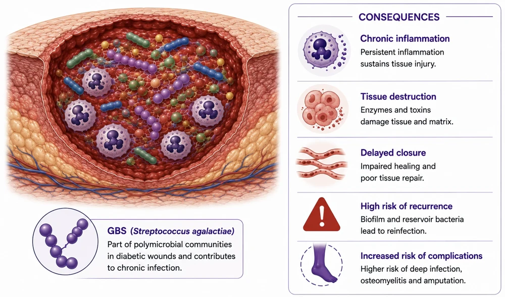
Diabetes mellitus affected 589 million adults worldwide in 2024 with a predicted global incidence to reach 853 million by 2050. Of these individuals, approximately one quarter will develop a diabetic foot ulcer (DFU) often resulting in non-healing wounds and chronic infection. DFUs frequently harbor complex polymicrobial communities dominated by bacterial species including GBS which is increasing isolated from diabetic wounds.
We developed a diabetic mouse model of GBS wound infection and are using a human cell line as well as human diabetic wound isolates to study host defense, and polymicrobial interactions in this niche.
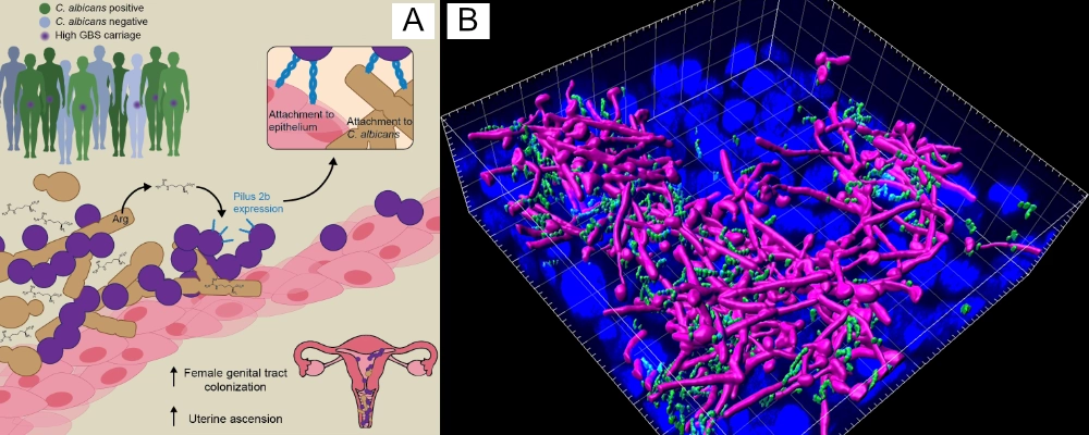
Figure (A) illustrates depicting bacterial and fungal interactions. Figure (B) is a three dimensional view of a GBS (green) - Candida albicans (fuchsia) biofilm on human vaginal epithelial cells.
We are interested in the ways that polymicrobial interactions can impact microbial physiology at the host interface. The interactions between bacteria and fungi have been understudied. Candida albicans (Ca) and GBS frequently colonize the vaginal tract, and we have recently demonstrated that Ca-GBS polymicrobial biofilms can protect GBS from antibiotic treatment and anchor GBS to the vaginal epithelial surface.
We have developed a new murine model to characterize the association between Ca and GBS in the vagina and seek to understand how these interkingdom interactions contribute to vaginal colonization and well as GBS persistence on other niches like the neonatal GI and diabetic wound.
Join the Lab
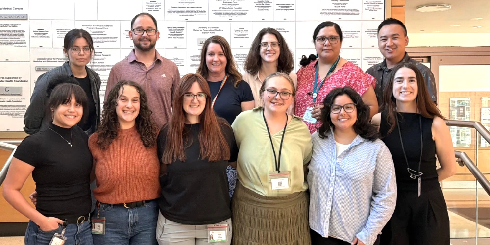
The Doran Lab provides excellent training, mentorship and research opportunities for students and trainees at all levels. We are always looking for highly motivated individuals that are excited about science!
Visiting Scientists and Post-doctoral Fellows interested in working in the lab are encouraged to contact Dr. Doran directly. Please include (1) a cover letter describing your career goals/scientific interests and highlighting your two most relevant publications and (2) a copy of your curriculum vitae including an updated list of peer-reviewed publications. Please arrange for three letters of recommendation to be sent to Dr. Doran via email. All postdocs will be expected to apply for their own funding.
Students interested in pursuing a MS or Ph.D., and interested in our research are encouraged to apply to the MIMS program or PhD Microbiology or Immunology graduate programs. Please contact us for further information about our research and opportunities.
Contact us
Kelly Doran, Ph.D.
Mail Stop 8333
Research Complex 1 North
12800 E. 19th Ave., Rm. P18-9105
Aurora, CO 80045
Office 303 724 3539
Lab 303 724 3539
FAX 303 724 42226


